

Retinal vasculature occlusion leads to carotid surgery
Submitted by: Kevin Stallard, OD
CASE SUMMARY
A 67-year-old female presented with the complaint of a blind spot in her right eye. She noticed it 2 days earlier and described it as gray and submarine-shaped. She stated that she was on cholesterol medicine and had experienced a stroke 2 years prior.
OUR PROCESS
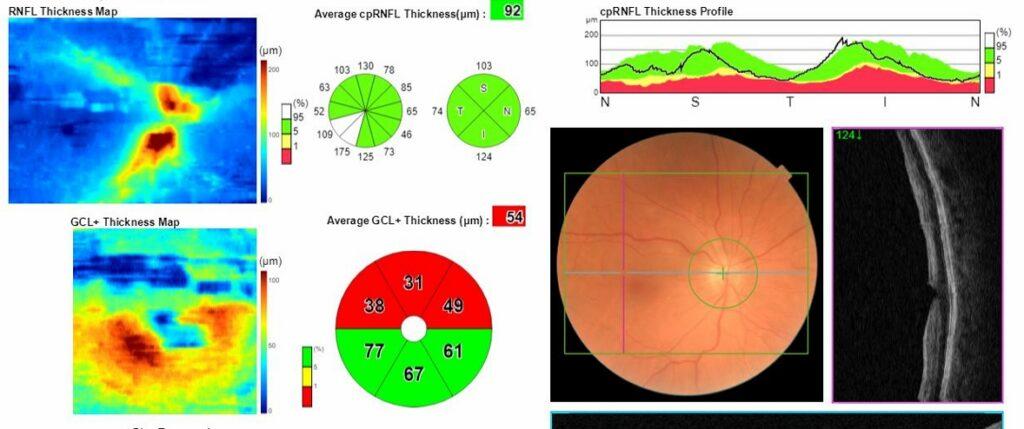
Entering unaided acuity was 20/60 right and 20/30- left. Pupils were equal, round, and responsive to light, with no APD. Motilities were full, and confrontation fields were normal except for a slight decrease inferiorly. Mild cataracts were noted in both eyes. Retinal examination revealed a branch retinal artery occlusion superiorly with visible Hollenhorst plaques in the right eye. There was ischemia noted superior and temporal to the optic nerve in the right eye. Mild epiretinal membranes were noted in both eyes. Left retina was otherwise normal. Retinal photography and an OCT of the retina were ordered and obtained.
TREATMENT
The patient was referred to the local emergency room for stroke assessment due to the artery occlusion and presence of emboli in the retina. After doing an evaluation, the local emergency room sent the patient to the University of Kentucky emergency department, and they ultimately tested her carotids. They found significant blockage, and she underwent carotid surgery the next day.
CONCLUSION
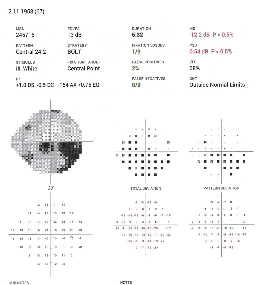
The patient returned to our office 2 months later for a follow-up. The retinal ischemia had improved, one of the original plaques was gone, and new plaque was noted in the temporal retina of the right eye. The right eye uncorrected acuity was 20/80 at this visit. Visual field testing revealed a decrease inferiorly, corresponding to the area of initial ischemia. She is continuing to be cared for by her medical doctor and will return to us in 3 months for repeat visual field and retinal testing.
NEVER IGNORE CHANGES IN YOUR VISION
Eye conditions can develop quietly, but early detection can make a significant difference in preserving your sight. If you’ve noticed changes in your vision, or it’s simply time for a comprehensive eye exam, our team is here to help.
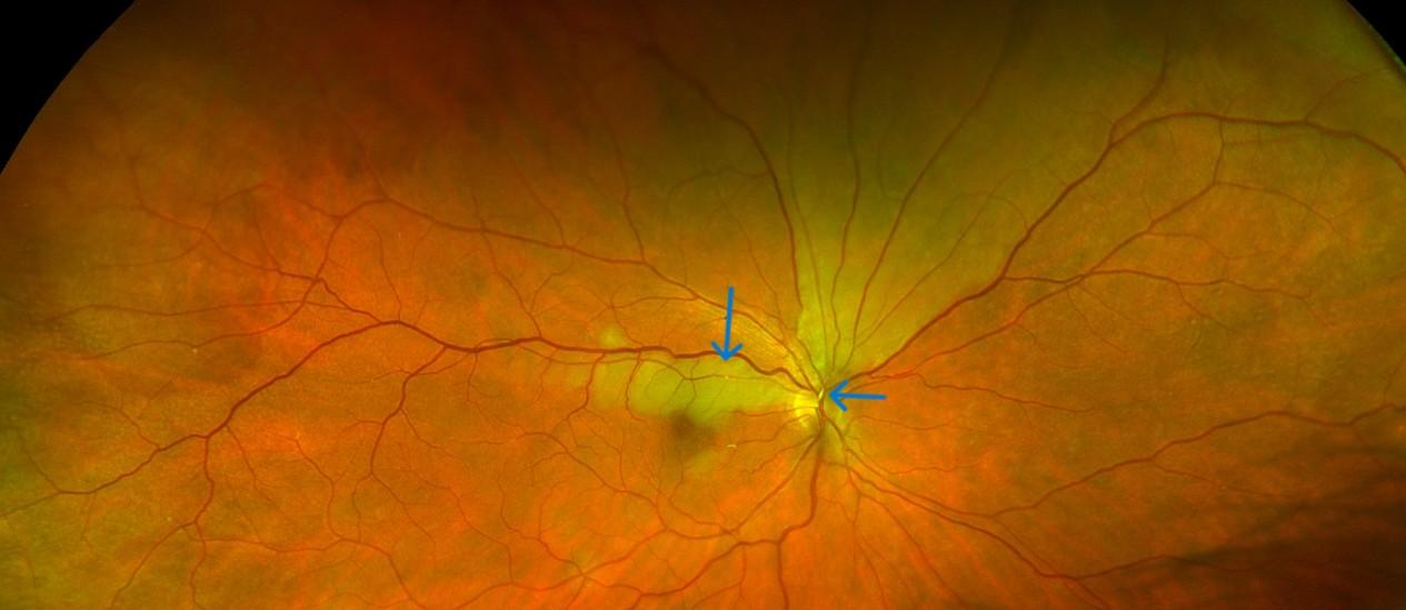
Initial Scan
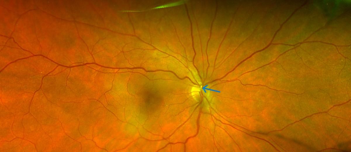
Post Treatment Scan